
Cardiac ablation is a procedure that can correct heart rhythm problems (arrhythmias). Ablation usually uses long, flexible tubes (catheters) inserted through a vein in your groin and threaded to your heart to correct structural problems in your heart that cause an arrhythmia.
Cardiac ablation works by scarring or destroying tissue in your heart that triggers an abnormal heart rhythm. In some cases, ablation prevents abnormal electrical signals from traveling through your heart and, thus, stops the arrhythmia.
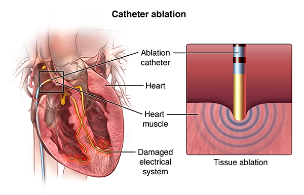
If you have been diagnosed with a heart arrhythmia-a problem with the rate or rhythm of your heartbeat-your doctor may recommend a procedure called catheter ablation to improve your condition.
Also known as a cardiac ablation or radiofrequency ablation, this procedure guides a tube into your heart to destroy small areas of heart tissue that may be causing your abnormal heartbeat.
Not everyone with a heart arrhythmia needs a catheter ablation. It's usually recommended for people with arrhythmias that can't be controlled by medication or with certain types of arrhythmia from the upper chambers of the heart. Less commonly, it may be recommended for people with arrhythmia that begins in the lower chambers of the heart.
Catheter ablation can take anywhere from 3 to 6 hours. The procedure is usually done in an electrophysiology lab or operating room where you will be monitored closely.
Before the procedure begins, you will be given intravenous medications to help you relax; some people even fall asleep. In some complex cases, you may be put to sleep by an anesthesiologist.
After the medication has taken effect, your doctor will numb an area on your arm, neck, or groin and make a small hole in your skin. Then, the doctor will guide a thin guide wire and 2 to 3 small catheters through blood vessels to your heart. In some cases, your doctor may place several catheters, which are used to help guide the procedures.
After the catheter has been placed correctly, electrodes at the end of the catheter are used to stimulate your heart and locate the problem areas that are causing the abnormal heart rhythm. Then, the doctor will use mild radiofrequency heat energy to destroy or "ablate" the problem area. This area is usually quite small, about one-fifth of an inch. Other types of ablation techniques may be used, such as cryoablation, which uses very cold temperatures to destroy the problem area. Your doctor will decide which type of ablation therapy is most appropriate for you. Once the tissue is destroyed, the abnormal electrical signals that created the arrhythmia can no longer be sent to the rest of the heart.
Most people do not feel pain during the procedure. You may sense mild discomfort in your chest. After the ablation is over, your doctor will remove the guide wire and catheter from your chest.
After the catheter ablation, you will probably need to lie still for 2 to 6 hours to decrease the risk of bleeding. Medical staff may apply pressure to the site where the catheter was inserted. Special machines will be used to monitor your heart as you recover. Some people can go home on the same day as the ablation, but others will stay in the hospital for one or more nights.
Recovery from catheter ablation is usually fairly straightforward. In the days after the procedure, you may experience mild symptoms such as an achy chest and discomfort or bruising in the area where the catheter was inserted. You might also notice skipped heartbeats or irregular heart rhythms. Most people can return to their normal activities within a few days.
Contact your doctor immediately if you have unusual pain or swelling, excessive bleeding, or consistent irregularities in your heartbeat.
Depending on the type of arrhythmia being treated, catheter ablation can have a success rate of more than 90%, but some people may need to have the procedure again or other treatments for heart arrhythmias. Your doctor may want you to remain on medications to help control your heartbeat.
After your catheter ablation, be sure to follow all instructions from your doctor, especially regarding follow-up visits, medication schedules, and safe levels of physical activity.
Online Medical Reviewer: Freeborn, Donna, PhD, CNM, FNP
Online Medical Reviewer: MMI board-certified, academically affiliated clinician
Date Last Reviewed: 2/12/2014
© 2000-2015 The StayWell Company, LLC. 780 Township Line Road, Yardley, PA 19067. All rights reserved. This information is not intended as a substitute for professional medical care. Always follow your healthcare professional's instructions.
Cardiac catheterizations are typically performed on patients who experience chest pain, shortness of breath or have had a stress test that revealed a possible cardiac problem. Known as a coronary angiography, a catheterization allows interventional cardiologists to see the arteries of the heart and locate blockages. They perform the procedure by injecting a contrast dye and then inserting a catheter into an artery and threading it through the blood vessels to the heart.
In addition to using catheterizations - both radial and transfemoral - for diagnoses, cardiologists can also treat blockages at the same time. They can perform an angioplasty - inserting a small balloon to widen the artery - or implant stents, small mesh devices to keep the artery walls open.
Cardiac catheterization is performed to further evaluate coronary artery disease, valvular heart disease, congestive heart failure, and/or certain congenital (present at birth) heart conditions, such as atrial septal defect or ventricular septal defect, when other less invasive types of diagnostic tests indicate the presence of one of these conditions.
In cardiac catheterization (often called cardiac cath), a very small hollow tube, or catheter, is advanced from a blood vessel in the groin or arm through the aorta into the heart. Once the catheter is in place, several diagnostic techniques may be used. The tip of the catheter can be placed into various parts of the heart to measure the pressures within the chambers. The catheter can be advanced into the coronary arteries and a contrast dye injected into the arteries.
The use of fluoroscopy (a special type of X-ray, similar to an X-ray "movie") assists the doctor in locating any blockages in the coronary arteries as the contrast dye moves through the arteries.
Angioplasty, percutaneous coronary intervention, and stenting may be done as part of, or following, a catheterization. Fractional flow reserve is a pressure management technique that is now commonly used in catheterization to determine the severity of an artery occlusion.
An additional technique called intravascular ultrasound (IVUS), a technique that uses a computer and a transducer that sends out ultrasonic sound waves to create images of the blood vessels, may be used during a cardiac cath. The use of IVUS provides direct visualization and measurement of the inside of the blood vessels and may assist the doctor in selecting the appropriate treatment needed in each particular situation.
A small sample of heart tissue (called a biopsy) may be obtained during the procedure to be examined later under the microscope for abnormalities.
The person will remain awake during the procedure, although a small amount of sedating medication will be given prior to the procedure to ensure the patient remains comfortable during the procedure.
Other related procedures that may be used to assess the heart include resting or exercise electrocardiogram (ECG or EKG), Holter monitor, signal-averaged ECG, chest X-ray, computed tomography (CT scan) of the chest, echocardiography, electrophysiological studies, myocardial perfusion scans, radionuclide angiography, magnetic resonance imaging (MRI) of the heart, and cardiac CT scan. Please see these procedures for additional information.
A cardiac catheterization may be performed to assist in the diagnosis of the following heart conditions:
A cardiac catheterization may also be performed if you have recently had an episode(s) of one or more of the following cardiac symptoms:
If a screening examination, such as an ECG or stress test suggests a possibility of a heart condition that needs to be explored further, a cardiac cath may be ordered by your doctor.
Other reasons for a cath procedure include evaluation of myocardial perfusion (blood flow to the heart muscle) if chest pain or angina occurs after the following:
There may be other reasons for your doctor to recommend a cardiac catheterization.
Possible risks associated with cardiac catheterization include, but are not limited to, the following:
You may want to ask your doctor about the amount of radiation used during the procedure and the risks related to your particular situation. It is a good idea to keep a record of your past history of radiation exposure, such as previous scans and other types of X-rays, so that you can inform your doctor. Risks associated with radiation exposure may be related to the cumulative number of X-ray examinations and/or treatments over a long period of time.
If you are pregnant or suspect that you may be pregnant, you should notify your doctor due to risk of injury to the fetus from a cardiac catheterization. Radiation exposure during pregnancy may lead to birth defects. If you are lactating, or breastfeeding, you should notify your doctor.
There is a risk for allergic reaction to the cath dye. Patients who are allergic to or sensitive to medications, contrast dye, iodine, or latex should notify their doctor. Also, patients with kidney failure or other kidney problems should notify their doctor.
For some patients, having to lie still on the cardiac catheterization table for the length of the procedure may cause some discomfort or pain.
There may be other risks depending on your specific medical condition. Be sure to discuss any concerns with your doctor prior to the procedure.
A cardiac catheterization may be performed on an outpatient basis or as part of your stay in a hospital. Procedures may vary depending on your condition and your doctor's practices.
Generally, a cardiac catheterization follows this process:
If the insertion site was in the arm, your arm will be kept elevated on pillows and kept straight by placing your arm in an arm guard (a plastic arm board designed to immobilize the elbow joint). In addition, a plastic band (works like a belt around the waist) may be secured around the arm near the insertion site. The band will be loosened at given intervals and removed at the appropriate time as determined by your doctor.
In the hospital
After the procedure, you may be taken to the recovery room for observation or returned to your hospital room. You will remain flat in bed for several hours after the procedure. A nurse will monitor your vital signs, the insertion site, and circulation/sensation in the affected leg or arm.
You should immediately inform your nurse if you feel any chest pain or tightness, or any other pain, as well as any feelings of warmth, bleeding, or pain at the insertion site in your leg or arm.
Bedrest may vary from two to six hours depending on your specific condition. If your physician placed a closure device, your bedrest may be of shorter duration.
In some cases, the sheath or introducer may be left in the insertion site. If so, the period of bedrest will be prolonged until the sheath is removed. After the sheath is removed, you may be given a light meal.
You may feel the urge to urinate frequently because of the effects of the contrast dye and increased fluids. You will need to use a bedpan or urinal while on bedrest so that your affected leg or arm will not be bent.
After the specified period of bed rest has been completed, you may get out of bed. The nurse will assist you the first time you get up, and will check your blood pressure while you are lying in bed, sitting, and standing. You should move slowly when getting up from the bed to avoid any dizziness from the long period of bedrest.
You may be given pain medication for pain or discomfort related to the insertion site or having to lie flat and still for a prolonged period.
You will be encouraged to drink water and other fluids to help flush the contrast dye from your body.
You may resume your usual diet after the procedure, unless your doctor decides otherwise.
When you have completed the recovery period, you may be discharged to your home unless your physician decides otherwise. Commonly, patients who undergo angioplasty or placement of a stent will spend the night in the hospital for careful observation. If this procedure was performed on an outpatient basis and a sedative was administered, you must have another person drive you home.
At home
Once at home, you should monitor the insertion site for bleeding, unusual pain, swelling, and abnormal discoloration or temperature change at or near the insertion site. A small bruise is normal. If you notice a constant or large amount of blood at the site that cannot be contained with a small dressing, notify your doctor.
If your doctor used a closure device for your insertion site, you will be given specific information regarding the type of closure device that was used and how to take care of the insertion site. There may be a small knot, or lump, under the skin at the site. This is normal. The knot should gradually disappear over a few weeks.
It will be important to keep the insertion site clean and dry. Your doctor will give you specific bathing instructions.
You may be advised not to participate in any strenuous activities for a period of time after the procedure. Your doctor will instruct you about when you can return to work and resume normal activities.
Notify your doctor to report any of the following:
Your doctor may give you additional or alternate instructions after the procedure, depending on your particular situation.
Online Medical Reviewer: Bass, Pat F. III, MD, MPH
Online Medical Reviewer: MMI board-certified, academically affiliated clinician
Date Last Reviewed: 10/31/2013
© The StayWell Company, LLC. 780 Township Line Road, Yardley, PA 19067. All rights reserved. This information is not intended as a substitute for professional medical care. Always follow your healthcare professional's instructions.
Cardioversion is a medical procedure that restores normal heart rhythm in people who have particular types of arrhythmias. It typically involves placing electrodes on the chest that send electric shocks to the heart and is usually done as an outpatient procedure. In some cases, physicians may prescribe medications to address the arrhythmias instead of performing a procedure.
An implantable cardioverter-defibrillator (ICD) is a battery-powered device placed under the skin that continuously monitors your heart rate. It is connected to your heart with thin wires and when it detects an abnormal heart rhythm it delivers an electric shock to restore your normal heartbeat.
An electrocardiogram (ECG/EKG) is a test that measures the electrical activity of the heart. It can be used to assess for heart attacks and arrhythmias. Its results can also suggest other disorders that affect heart function.
An echocardiogram uses sound waves to produce images of your heart. This commonly used test allows your doctor to see your heart beating and pumping blood. Your doctor can use the images from an echocardiogram to identify heart disease.
Depending on what information your doctor needs, you may have one of several types of echocardiograms. Each type of echocardiogram has few, if any, risks involved.
A nuclear stress test measures blood flow to your heart at rest and while your heart is working harder as a result of exertion or medication. The test provides images that can show areas of low blood flow through the heart and damaged heart muscle.
The test usually involves taking two sets of images of your heart — one while you're at rest and another after you heart is stressed, either by exercise or medication.
You may be given a nuclear stress test, which involves injecting a radioactive dye into your bloodstream, if your doctor suspects you have coronary artery disease or if a routine stress test didn't pinpoint the cause of symptoms such as chest pain or shortness of breath. A nuclear stress test may also be used to guide your treatment if you've been diagnosed with a heart condition.
A pacemaker insertion is the implantation of a small electronic device that is usually placed in the chest (just below the collarbone) to help regulate slow electrical problems with the heart. A pacemaker may be recommended to ensure that the heartbeat does not slow to a dangerously low rate.
Preventing heart disease is possible, by recognizing your symptoms and risk factors early.
Our expert cardiologists offer an integrated approach to care, including:
A transesophageal echocardiogram (TEE) is a procedure performed to evaluate your heart and the surrounding structures. The esophagus lies directly behind the heart, allowing for better quality images generally than those obtained from an echocardiogram from the chest wall. It is especially valuable in patients with valve replacements, history of a stroke, or when adequate images cannot be obtained from the chest wall.
To perform a TEE, a small flexible tube containing a small transducer is passed into the esophagus. Prior to the test, the back of your throat will be numbed first with a medication you will swallow, followed by another medication that will be sprayed into your mouth. You will then be given medication intravenously for sedation. All of this is done to make the procedure more comfortable for you.
A vascular ultrasound is the general term for a non-invasive painless test that uses high-frequency sound waves to image blood vessels including arteries and veins.
Lower extremity venous ultrasound is typically performed if a clot in the vein (deep venous thrombosis or DVT) is suspected. The veins in the legs are compressed and the blood flow is assessed to make sure the vein is not clogged. This test is also used to look for chronic venous insufficiency, or leaky valves in the veins which may cause swelling or edema.
Lower extremity arterial ultrasound may be performed in patients with peripheral artery disease (PAD), particularly for planning an endovascular procedure or surgery. It is also used after the procedure to monitor stents and grafts for signs of the blockage returning ("restenosis"). If a hematoma develops after a catheterization procedure, arterial ultrasound is also used to check the integrity of the arteries and veins in the groin.
A carotid ultrasound is a non-invasive, painless test that uses high-frequency sound waves to image the neck arteries.
Atherosclerosis may occur in the blood vessels in the neck (the "carotid arteries") which supply blood to the brain. This condition is called carotid stenosis and is typically diagnosed using carotid ultrasound. This technique allows us to look for atherosclerotic plaque and to assess whether this plaque is interfering with blood flow to the brain. As the artery narrows, the velocity of the blood flow increases; ultrasound allows us to measure the speed of the blood flow in order to estimate the degree of blockage.
Your doctor may order a carotid ultrasound if a blockage is suspected based on listening to your neck or based on your cardiovascular risk profile. This test should also be performed if you have had a stroke or a Transient Ischemic Attack (TIA).
An ultrasound of the abdominal aorta is a non-invasive, painless test that uses high-frequency sound waves to image the "aorta," the main blood vessel leading away from the heart.
When the walls of the abdominal aorta become weak, they may balloon outward If the aorta reaches over 3 centimeters in diameter, it is then called an abdominal aortic aneurysm (AAA). As the aneurysm gets larger, the risk of rupture increases.
Ultrasound imaging of the aorta is useful for measuring its size to screen for AAA. Screening is particularly recommended for men over the age of 60 who have ever smoked and for anyone with a family history of AAA. In addition to screening, ultrasound is also a useful tool after the diagnosis of AAA to monitor its size on a regular basis to see if it needs to be repaired.
Vascular studies are a noninvasive (the skin is not pierced) procedure used to assess the blood flow in arteries and veins. A transducer (like a microphone) sends out ultrasonic sound waves at a frequency too high to be heard. When the transducer is placed on the skin at certain locations and angles, the ultrasonic sound waves move through the skin and other body tissues to the blood vessels, where the waves echo off of the blood cells. The transducer picks up the reflected waves and sends them to an amplifier, which makes the ultrasonic sound waves audible.
Vascular studies can utilize one of these special types of ultrasound technology, as listed below:
Doppler ultrasound: This Doppler technique is used to measure and assess the flow of blood through the blood vessels. The amount of blood pumped with each beat is an indication of the size of a vessel's opening. Also, Doppler can detect abnormal blood flow within a vessel, which can indicate a blockage caused by a blood clot, a plaque, or inflammation.
Color Doppler: Color Doppler is an enhanced form of Doppler ultrasound technology. With color Doppler, different colors are used to designate the direction of blood flow. This simplifies the interpretation of the Doppler technique.
To assess blood flow in the limbs, pulse volume recordings (PVRs) may be performed. Blood pressure cuffs are inflated on the limb and blood pressure in the limb is measured using the Doppler transducer.
To assess the carotid arteries in the neck, a carotid duplex scan may be performed. This type of Doppler examination provides a 2-dimensional (2D) image of the arteries so that the structure of the arteries and location of an occlusion can be determined, as well as the degree of blood flow.
A carotid artery duplex scan is a type of vascular ultrasound study done to assess occlusion (blockage) or stenosis (narrowing) of the carotid arteries of the neck and/or the branches of the carotid artery. Plaque (a buildup of fatty materials), a thrombus (blood clot), and other substances in the blood stream may cause a disturbance in the blood flow through the carotid arteries.
Other related procedures that may be used to assess the heart and circulatory system include resting and exercise electrocardiogram (ECG or EKG), Holter monitor, signal-averaged ECG, cardiac catheterization, chest X-ray, computed tomography (CT scan) of the chest, electrophysiological studies, magnetic resonance imaging (MRI) of the heart, myocardial perfusion scans, radionuclide angiography, and ultrafast CT scan. Please see these procedures for additional information.
The arteries bring oxygen and other nutrients to the cells of the body. The veins take away the blood after the cells have taken in the oxygen and nutrients and given up their waste products, such as carbon dioxide. If blood flow is decreased to any part of the body, that area does not get enough oxygen and nutrients and is unable to get rid of its waste products adequately.
Decreased blood flow can occur in the arteries and veins anywhere in the body, such as the neck and brain. When the neck arteries (carotid arteries) become occluded, symptoms such as dizziness, confusion, drowsiness, headache, and/or a brief loss of ability to speak or move, may be the early warning signs of a possible stroke (brain attack). More severe symptoms, such as sudden sharp headache, loss of vision in one eye, sudden loss of ability to move arms, legs, or one side of the body, sudden forceful vomiting, or sudden decreased level of consciousness may mean that a stroke is imminent.
Some conditions which may affect blood flow include, but are not limited to, the following:
If the doctor suspects that a person may have decreased blood flow somewhere in the peripheral (arms, legs, and/or neck) circulation, vascular studies may be performed.
Evaluation of signs and symptoms which may suggest decreased blood flow in the arteries and/or veins of the neck, legs, or arms
Evaluation of previous procedures that were performed to restore blood flow to an area
Evaluation of a vascular dialysis device, such as an A-V fistula in the arm
There may be other reasons for your doctor to recommend a vascular study.
There is no radiation used and generally no discomfort from the application of the ultrasound transducer to the skin.
For some patients, having to lie still on the examination table for the length of the procedure may cause some discomfort or pain.
There may be other risks depending on your specific medical condition. Be sure to discuss any concerns with your doctor prior to the procedure.
Certain factors or conditions may interfere with a vascular study. These factors include, but are not limited to, the following:
A vascular study may be performed on an outpatient basis or as part of your stay in a hospital. Procedures may vary depending on your condition and your doctor's practices.
Generally, a vascular study follows this process:
Online Medical Reviewer: Bass, Pat F. III, MD, MPH
Online Medical Reviewer: MMI board-certified, academically affiliated clinician
Date Last Reviewed: 10/31/2013
© The StayWell Company, LLC. 780 Township Line Road, Yardley, PA 19067. All rights reserved. This information is not intended as a substitute for professional medical care. Always follow your healthcare professional's instructions.
954 Teaneck Road, Teaneck, NJ 07666
P: 201-833-2300 | F: 201-833-7600
Meet Our Physicians
Stephen Angeli, MD
Gerard Eichman, MD
Tariqshah Syed, MD
David Wild, MD
Shalin P. Desai, MD

718 Teaneck Road, Teaneck, NJ 07666
P: 1-877-HOLY-NAME (465-9626)
This information is not intended as a substitute for professional medical care. Always follow your healthcare professional's instructions.
Copyright © 2026 Cardiovascular Specialists of North Jersey, All rights reserved.